Oral Diagnosis and Radiology
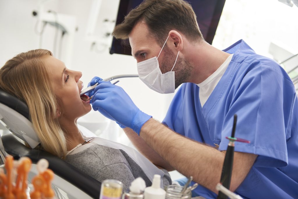
Oral Diagnosis and Radiology, which literally means intraoral diagnosis; It is the first clinic that patients visit before starting their treatment in dentistry faculties, private hospitals or dental clinics. By specialist dentists in the Oral Diagnosis and Radiology clinic; Intraoral examination is performed and panoramic x-ray is taken showing the structure of the teeth and jaw. After this stage, a patient-specific treatment plan is drawn up and treatment is provided in the relevant department. However, problems such as decayed teeth, impacted teeth or receding gums are not only seen in the mouth. There may also be diseases seen in the soft tissues on the cheek, palate and jaw bones. In the Oral Diagnosis and Radiology clinic, systemic diseases such as diabetes and Behçet’s diseases, which are found elsewhere in the body but have symptoms in the oral tissues, and intraoral tissue diseases such as everyone and aphtha are also diagnosed and treated.

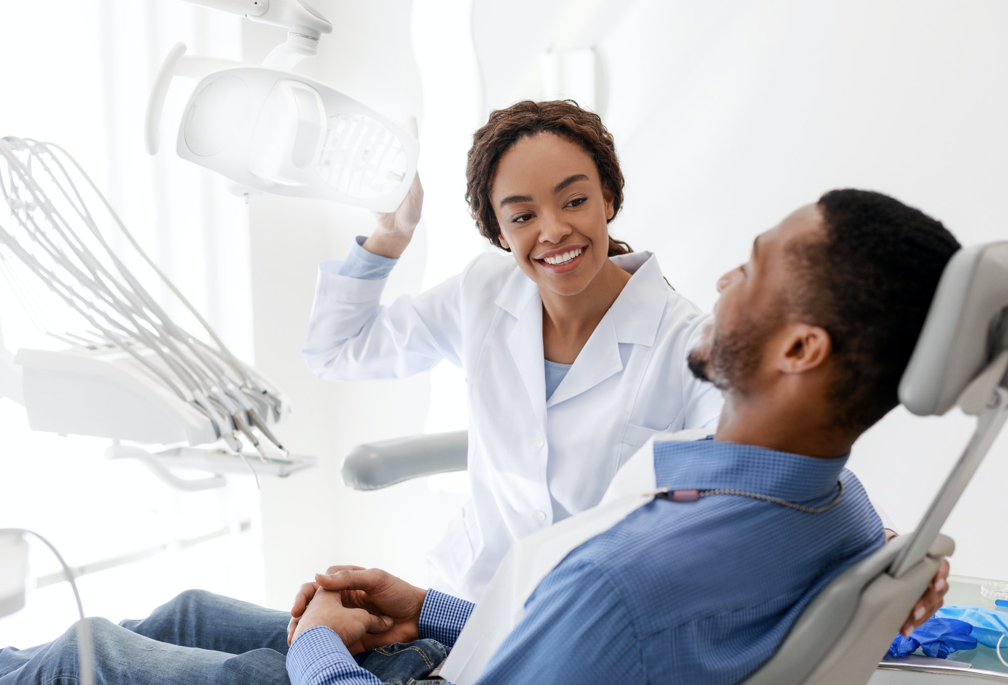
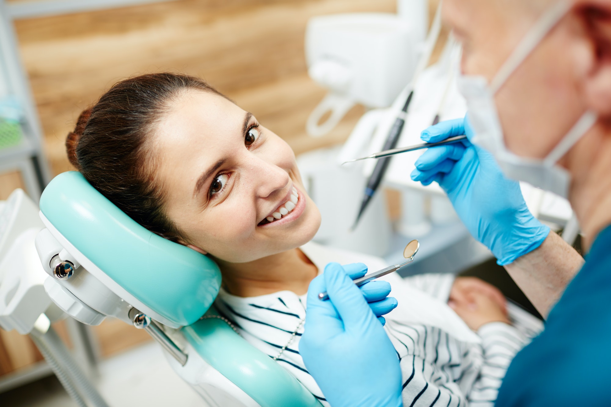
A successful dental treatment can only be done if a good diagnosis is made, as in all diseases. From this point of view, Oral Diagnosis actually fulfills a very important task. Intraoral examination; It covers all soft tissues in the face, jaw and jaw joint, lips, palate, tongue, nose and neck areas. In addition, panoramic x-rays should be taken in order to clearly see the tooth alignment and roots. Gingival recessions, bacterial plaques, dental calculus, fistulas, caries, overlapping teeth and jaw closures in the mouth are evaluated. At this stage, teeth that are not yet painful or gums that have just begun to recede should also be evaluated and conservative treatment should be applied. In addition, patients who come for treatment should be asked about their existing heart, vascular, blood pressure, allergy, asthma, diabetes, hormonal disorders and the drugs they regularly use. The existence of such disorders will change both the treatment method and the drugs to be used in the treatment. Dental treatment will be planned by taking the patient’s entire history in the Oral Diagnosis and Radiology clinic. The patient’s expectations from dental treatment are learned and the possible results are explained to the patient in detail and treatment is recommended.
One of the most effective methods of helping dental treatments is; radiological examinations. These examinations are; dental tomography, intraoral and extraoral x-rays, panoramic x-rays and periapical x-rays. Of these, in dental tomography; A large number of bone and soft tissue images are taken from axial, sagittal and coronal angles that cannot be seen with normal x-rays. These images are also reconstructed with advanced special software, allowing the region to be imaged to be viewed from any angle and from any direction. These sections, which give all the details and accurate measurements necessary for diagnosis, will also guide the treatment. Intraoral and extraoral x-rays are periapical x-rays that can be easily taken in every dentist’s office and show one or more teeth. In these X-ray images, along with the tooth, root, bone loss, caries, tooth and tooth root infections can be easily seen.
Panoramic x-rays are x-rays that show all the teeth in the mouth, impacted teeth, jaw bone structure, cavities in the mouth and jaw joints. It is used in oral examination for control purposes, extraction of impacted teeth, detection of cysts or tumors, and surgical applications such as applying one or more implants. Periapical x-rays are; It is the method used to obtain more detailed images of dental disorders detected in panoramic x-rays. A superior image is obtained compared to panoramic x-rays in terms of detailed image acquisition. However, the exposure rate in panoramic x-rays is much lower than in periapical x-rays.

1. Implant
2. Dental Fillings

3. Tooth Extraction
4. Whitening